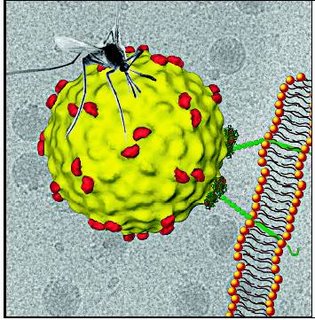
In a scientific breakthrough, an international team of virologists led by a Howard Hughes Medical Institute scholar in Argentina, Andrea Gamarnik, has identified the mechanism by which the dengue virus replicates its genetic code inside the infected cell, thus triggering the fatal Dengue Hemorrhagic Fever (DHF).
With no specific medicine yet having been found for the treatment of the disease, this finding will now help scientists to understand how the virus propagates and ultimately find strategies to control the disease.
The dengue virus belongs to a family of viruses know as Flaviviruses. This group of viruses also cause diseases like Yellow Fever, West Nile and Encephalitis. Till now, little was known about the mechanism of Flavivirus RNA replication. Because the essential RNA signals found for dengue virus replication are also present in other Flavivirus genomes, it is likely that the findings can be extrapolated to other members of the family.
In India, DHF is the leading cause of hospitalisation and death among children. Dengue virus was first isolated in India in 1945. The number of cases range between 9,000 and 12,000 per annum. In Kolkata, over 32 people died of DHF last year.
Gamarnik and his team explained the mechanism by which dengue virus replicates its genetic code inside the infected cell. Dengue and other Flaviviruses belong to a group whose genetic material is composed of Ribonucleic Acid (RNA). RNA contains the code to produce the viral proteins, especially a protein called RNA Polymerase. The polymerase is the enzyme responsible for copying the RNA to produce large amounts of this molecule, which is essential for viral multiplication.
Gamarnik said: “A rapid amplification of the viral RNA from one single molecule to tens of thousands of copies occurs in only a few hours after infection. But when the polymerase is ready to start copying, the viral RNA is surrounded by millions of cellular RNA molecules. In our lab we identified a signal in the viral RNA that attracts the polymerase. The polymerase starts copying the molecule from the end on the right.”
He added: “How does the polymerase jump from one end to the other was a puzzle. Using biochemical and genetic tools together with atomic force microscopy, we found that the viral RNA forms circles bringing together different ends of the RNA. This explained how the polymerase copies the RNA. “As shown for other viruses, such as HIV, the viral polymerases are effective targets for development of antiviral treatments. Therefore, knowing how the dengue virus polymerase works, how is its 3D structure and how it recognises the viral RNA are fundamental questions that will help us develop antiviral solutions.”
The finding will be published in the August 15 issue of the journal Genes and Development. The team, which includes scientists Claudia Filomatori, Maria Lodeiro, Diego Alvarez, Marcelo Samsa and Lma Pietrasanta, has been working on the research for the past eight years. The team now wants to find out how the 3D structure of the polymerase bind to the viral RNA and whether scientists can interfere with viral RNA synthesis to stop dengue virus infection.
THE KEY TO THE CODE
- The above image depicts the Velcro-like protein on a cell's surface just after it attached to the dengue virus, a linkup thought to initiate the early stages of infection.
- The dengue virus belongs to the Flavivirus family of viruses. This group also causes Yellow Fever, West Nile and Encephalitis.
- RNA contains the code to produce the viral proteins, called RNA Polymerase. This is the enzyme responsible for copying the RNA to produce large amounts of this molecule, which is essential for viral multiplication.
- When the polymerase is ready to start copying, the viral RNA is surrounded by millions of cellular RNA molecules.
- Scientists identified a signal in the viral RNA that attracts the polymerase.

